

Sjögren’s Disease (SjD) is a systemic autoimmune disease characterized by lymphocytic infiltration and dysfunction of the exocrine glands, notably the lacrimal and salivary glands. The oral consequences include salivary gland dysfunction, altered salivary compositions, and loss of protective effects from saliva such as buffering capacity, antimicrobial capabilities, and clearance of bacteria and food debris.1 The disease frequently goes unrecognized due to its invisibility since patients appear to look “relatively normal” yet suffer from numerous debilitating systemic effects. Patient complaints of dry mouth often provoke dismissive comments such as “There, there dear, drink water” without the realization that water is not substantive, it does not remain in the mouth, and that the little saliva that is present gets swallowed along with the water. This subsequently makes the kidneys work overtime leading to an electrolyte imbalance and interrupted sleep due to frequent nocturnal bathroom visits.
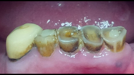
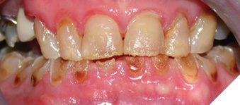
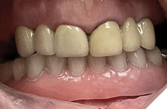
Patients frequently present with difficulty swallowing, speaking, tasting, eating, and chewing; a dry oral mucosa that is prone to ulceration; a dysbiosis created in the oral microflora; an increase in aciduric/acidogenic genera; and a more caries-susceptible environment.2 The associated detrimental effects of dry mouth, whether patient-perceived (xerostomia) or objectively measured (salivary gland dysfunction), can lead to a diagnosis of hyposalivation accompanied by numerous oral manifestations: rampant tooth decay, frequent deterioration of dental restorations, increased tooth loss, and oral candidiasis (Fig. 1-3). Although clinical guidelines for SjD patients have recommended fluoride for caries management,3 evidence of its effectiveness is sparse.4,5 Furthermore, not only is the oral environment drier and more acidic, and as a result not optimal for the usual culprit of oral decay, Strep. mutans, but the unique cervically located and smooth surface decay pattern in SjD patients does not conform to those in otherwise healthy salivators. This suggests that species other than Strep. mutans may be held suspect. We previously reported6,7 through real-time PCR that Actinomyces naeslundii, Scardovia wiggsiae, Lactobacillus fermentum, and Lactobacillus acidophilus were all significantly elevated in oral plaque obtained from SjD patients and that Scardovia wiggsiae, which is associated with Severe Early Childhood Caries (SECC)8 is also an indicator species in SjD.
Fig. 1A
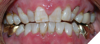
Fig. 1B
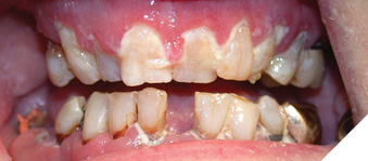
Fig. 1C
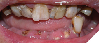
Fig. 2
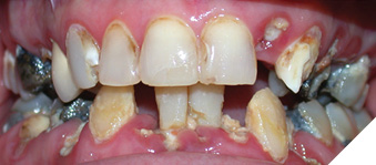
Fig. 3
Despite frequent dental visits and vigilant oral hygiene practices, including repeated applications of high fluoride-containing products,9,10 SjD patients have been observed to exhibit persistent tooth decay. This suggests that although fluoride is present to allow for remineralization of tooth structure, the unique decay-causing bacteria associated with SjD persist.
In recent years, silver diamine fluoride (SDF) has been approved for use in North America as minimally invasive treatment for dentin hypersensitivity and carious lesions.11 Evidence has indicated that SDF is 89% more effective in controlling/arresting caries progression than other treatments.12,13 The silver ions exert broad-spectrum antimicrobial effects through mechanisms such as disrupting bacterial cell membranes and/or walls, inhibiting enzyme activity and inactivating bacterial nucleic acids. The fluoride enhances remineralization and prevents further demineralization of tooth structure. Numerous reports have indicated that SDF is highly effective in arresting and preventing carious lesions, particularly in pediatric patients suffering from SECC14-16 (Fig. 4). 17
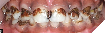
To date, there are no clinical trials that have explored whether SDF may confer similar oral health benefits for dry mouth patients, and particularly SjD patients, who share a similar decay pattern (Fig. 4A) 17 to those with SECC. The objective of the current clinical study was to determine the effectiveness of SDF in decreasing oral decay in patients with SjD.
Fig. 4A
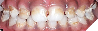
Fig. 4B
SDF was first licensed for use in Canada early in 2017 under the name Advantage Arrest (Oral Science). Since that time, there have been evidence-based guidelines18 and extensive evidence in support of the use of SDF to arrest caries19 with indications for treatment of dental caries in cases of extreme caries risk (Xerostomia or Severe Early Childhood Caries).
Clinical study
Participants previously diagnosed with SjD were recruited into the clinical study. Each participant was given written/verbal information about the advantages/disadvantages of SDF application.11 Informed patient consent was obtained,11 and the presence of carious lesions and location of patient-reported sensitivity were noted.
Phase 1 of the study focused on only posterior teeth. It consisted of cotton roll isolation, followed by SDF application to all surfaces of non-crowned posterior teeth or to gingival margins of crowned teeth in contralateral quadrants in opposing arches for two minutes. Areas of black staining were noted. Two to six weeks later, changes to the previously SDF-treated areas with respect to hardness and in sensitivity were noted. SDF was then similarly applied to posterior teeth in the remaining two quadrants. Re-examination and SDF re-application occurred three months following the first SDF application and at subsequent appointments at three-month intervals for up to four years.
Phase 2 of the study followed a similar procedure but included SDF application to remaining non-aesthetic areas such as the lingual/palatal surfaces of anterior teeth. Where appropriate, cavitated lesions arrested by SDF were subsequently restored through minimally invasive techniques with composite resin (CR) or glass ionomer (GI) (Figs. 5-7). In cases where teeth were deemed non-restorable but useful to maintain bone levels, the roots were SDF-treated (Fig. 8A) and retained to act as overdenture abutment teeth (Figs. 8B & C). Patient satisfaction questionnaires were completed. Of note is that those SjD patients with the driest of mouths, often commented that the SDF stung their gums. This was short-lived and disappeared five to ten minutes following the application. Any saliva present initially also became a white opaque fluid for a few minutes, as opposed to a colourless fluid. Also short-lived was that the application of SDF frequently stimulated the release of saliva, possibly in response to the bitter metallic taste.
Fig. 5A
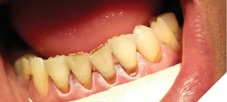
Fig. 5B
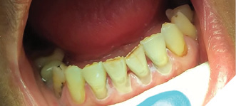
Fig. 6A
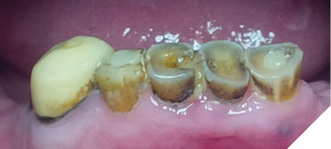
Fig. 6B
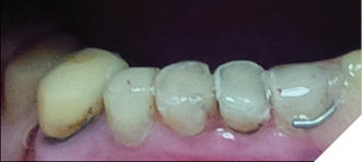
Fig. 7A
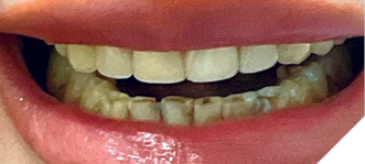
Fig. 7B
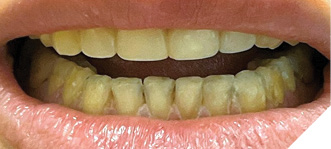
Fig. 8A
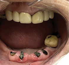
Fig. 8B
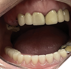
Fig. 8C
Results
To date, over eighty-five SjD patients have participated in this ongoing “black magic” study. In SjD participants, SDF stained only those areas of enamel and/or dentin where softness had initially been detected or at borders of previously placed CR restorations. The rate of decay significantly decreased following the initial SDF application. This continued with three- to six-month reapplication and remained over the testing period for up to four years. Previously noted lesions were stable. Patients reported a cessation of sensitivity as well as an increase in self-esteem, self-confidence, and satisfaction with restored teeth at minimal expense.
Conclusion
The significance of our findings indicates that the clinical use of SDF in SjD patients can effectively reduce/arrest caries progression which had previously been elusive. This treatment, however, is neither guaranteed nor static and is subject to fluctuations in severity of SjD symptoms. What it does provide is a team approach with the goal of improved oral health and a durable, inexpensive, aesthetic option for the SjD patient. 
Oral Health welcomes this original article.
References
- Humphrey SP, Williamson RT. A review of saliva: normal composition, flow, and function. J Prosthet Dent. 2001 Feb;85(2):162-9. doi: 10.1067/mpr.2001.113778. PMID: 11208206.
- Proctor DM, Fukuyama JA, Loomer PM, Armitage GC, Lee SA, Davis NM, Ryder MI, Holmes SP, Relman DA. A spatial gradient of bacterial diversity in the human oral cavity shaped by salivary flow. Nat Commun. 2018 Feb 14;9(1):681. doi: 10.1038/s41467-018-02900-1. PMID: 29445174; PMCID: PMC5813034.
- Zero DT, Brennan MT, Daniels TE, Papas A, Stewart C, Pinto A, Al-Hashimi I, Navazesh M, Rhodus N, Sciubba J, Singh M, Wu AJ, Frantsve-Hawley J, Tracy S, Fox PC, Ford TL, Cohen S, Vivino FB, Hammitt KM; Sjögren’s Syndrome Foundation Clinical Practice Guidelines Committee. Clinical practice guidelines for oral management of Sjögren disease: Dental caries prevention. J Am Dent Assoc. 2016 Apr;147(4):295-305. doi: 10.1016/j.adaj.2015.11.008. Epub 2016 Jan 5. PMID: 26762707.
- Xin W, Leung KC, Lo EC, Mok MY, Leung MH. A randomized, double-blind, placebo-controlled clinical trial of fluoride varnish in preventing dental caries of Sjögren’s syndrome patients. BMC Oral Health. 2016 Sep 23;16(1):102. doi: 10.1186/s12903-016-0296-7. Erratum in: BMC Oral Health. 2017 Mar 17;17(1):63. doi: 10.1186/s12903-017-0350-0. PMID: 27664129; PMCID: PMC5034648.
- Xin W, Leung KC, Lo EC, Mok MY, Leung MH. Erratum to: A randomized, double-blind, placebo-controlled clinical trial of fluoride varnish in preventing dental caries of Sjögren’s syndrome patients. BMC Oral Health. 2017 Mar 17;17(1):63. doi: 10.1186/s12903-017-0350-0. Erratum for: BMC Oral Health. 2016 Sep 23;16(1):102. doi: 10.1186/s12903-016-0296-7. PMID: 28314382; PMCID: PMC5357329.
- Trifonov, VH, Hall, MW, Cordova, M, Neufeld, JD, Laing, LP, Tennenbaum, HC, Beiko, RG, and Senadheera, DB. February 2016. “Harnessing the Oral Microbiome to Develop a Novel Plaque-Based Pathogen Monitoring Method for Sjögren’s Syndrome”. Oral presentation. Faculty of Dentistry Research Day.
- Laing Gibbard, LP, Trifonov, VH, Hall, MW, Cordova, M, Neufeld, JD, Tennenbaum, HC, Beiko, RG, and Senadheera, DB. September 2016. Strep. mutans: Partner in Crime for Sjögren’s Dental Decay but not the Ringleader. Biomarkers and Therapeutic Targets in Sjögren’s Conference. Oklahoma City Oklahoma USA. Poster presentation.
- Tanner AC, Mathney JM, Kent RL, Chalmers NI, Hughes CV, Loo CY, Pradhan N, Kanasi E, Hwang J, Dahlan MA, Papadopolou E, Dewhirst FE. Cultivable anaerobic microbiota of severe early childhood caries. J Clin Microbiol. 2011 Apr;49(4):1464-74. doi: 10.1128/JCM.02427-10. Epub 2011 Feb 2. PMID: 21289150; PMCID: PMC3122858.
- Pedersen AM, Bardow A, Nauntofte B. Salivary changes and dental caries as potential oral markers of autoimmune salivary gland dysfunction in primary Sjogren’s syndrome. BMC Clin Pathol. 2005 Mar 1;5(1):4. doi: 10.1186/1472-6890-5-4. PMID: 15740617; PMCID: PMC554998.
- AlmståhI A, Wikström M, Stenberg I, Jakobsson A, Fagerberg-Mohlin B. Oral microbiota associated with hyposalivation of different origins. Oral Microbiol Immunol. 2003 Feb;18(1):1-8. doi: 10.1034/j.1399-302x.2003.180101.x. PMID: 12588452.
- Horst JA, Ellenikiotis H, Milgrom PL. UCSF Protocol for Caries Arrest Using Silver Diamine Fluoride: Rationale, Indications and Consent. J Calif Dent Assoc. 2016 Jan;44(1):16-28. PMID: 26897901; PMCID: PMC4778976.
- Chibinski AC, Wambier LM, Feltrin J, Loguercio AD, Wambier DS, Reis A. Silver Diamine Fluoride Has Efficacy in Controlling Caries Progression in Primary Teeth: A Systematic Review and Meta-Analysis. Caries Res. 2017;51(5):527-541. doi: 10.1159/000478668. Epub 2017 Oct 4. PMID: 28972954.
- Oliveira BH, Cunha-Cruz J, Rajendra A, Niederman R. Controlling caries in exposed root surfaces with silver diamine fluoride: A systematic review with meta-analysis. J Am Dent Assoc. 2018 Aug;149(8):671-679.e1. doi: 10.1016/j.adaj.2018.03.028. Epub 2018 May 24. PMID: 29805039; PMCID: PMC6064675.
- Mei ML, Lo ECM, Chu CH. Arresting Dentine Caries with Silver Diamine Fluoride: What’s Behind It? J Dent Res. 2018 Jul;97(7):751-758. doi: 10.1177/0022034518774783. Epub 2018 May 16. PMID: 29768975.
- Chu CH, Mei L, Seneviratne CJ, Lo EC. Effects of silver diamine fluoride on dentine carious lesions induced by Streptococcus mutans and Actinomyces naeslundii biofilms. Int J Paediatr Dent. 2012 Jan;22(1):2-10. doi: 10.1111/j.1365-263X.2011.01149.x. Epub 2011 Jun 27. PMID: 21702854.
- Mei ML, Chu CH, Low KH, Che CM, Lo EC. Caries arresting effect of silver diamine fluoride on dentine carious lesion with S. mutans and L. acidophilus dual-species cariogenic biofilm. Med Oral Patol Oral Cir Bucal. 2013 Nov 1;18(6):e824-31. doi: 10.4317/medoral.18831. PMID: 23722131; PMCID: PMC3854072.
- Nuvvula S, Mallineni SK. Silver Diamine Fluoride in Pediatric Dentistry. J South Asian Assoc Pediatr Dent 2019;2(2):73–80.
- Slayton RL, Urquhart O, Araujo MWB, Fontana M, Guzmán-Armstrong S, Nascimento MM, Nový BB, Tinanoff N, Weyant RJ, Wolff MS, Young DA, Zero DT, Tampi MP, Pilcher L, Banfield L, Carrasco-Labra A. Evidence-based clinical practice guideline on nonrestorative treatments for carious lesions: A report from the American Dental Association. J Am Dent Assoc. 2018 Oct;149(10):837-849.e19. doi: 10.1016/j.adaj.2018.07.002. PMID: 30261951.
- Seifo N, Cassie H, Radford JR, Innes NPT. Silver diamine fluoride for managing carious lesions: an umbrella review. BMC Oral Health. 2019 Jul 12;19(1):145. doi: 10.1186/s12903-019-0830-5. PMID: 31299955; PMCID: PMC6626340.
About the Authors

Dr. Leslie P. Laing is a clinical associate in the Department of Prosthodontics at the Faculty of Dentistry University of Toronto; a Prosthodontist and Dry Mouth Specialist at Toronto Public Health Dental and Oral Care Clinics; is in private practices devoted to implant and Prosthodontic dentistry and relief of dry mouth in Toronto, Mississauga and Brockville; and is the President of the Sjögren’s Society of Canada.

Dr. Javid A. Karim graduated with a DDS from the University of Toronto in 2022. In 2024, he completed a 2-year General Practice Residency from the University of Rochester, Eastman Institute for Oral Health. He currently practices general dentistry in Toronto.