
Since the carious process is initiated and mediated by bacteria and their by products, mechanical removal of the caries and a thorough debridement of the cavity preparation is the accepted clinical norm. 1 When using dental sealants the possibility of sealing in active decay has been extensively debated in the dental literature. Numerous studies have shown that when one seals in caries (and the causative bacterial load), the caries process is arrested, there is a decrease in micro-organisms with time, or the bacteria are completely eliminated as the lesion remains totally sealed from the oral environment. 2,3
The concern of course, from a diagnostic perspective, is that it is often impossible to see microleakage under dental sealants especially if they are tooth coloured. It is therefore imperative when placing sealants, that the manufacturer’s protocol is strictly followed, preferably using an acid etch material,
which has a surfactant in it (Ultra-Etch Ultradent) (Fig. 1) to maximize penetration, etching and cleaning of the enamel defects. As well the clinician should use a penetrating sealant material like Ultraseal XT (Ultradent) (Fig. 2) which has one of the highest retention rates ever documented. 4
Glass ionomer cements have been used in dentistry for a long period of time. As a restorative material, the release of fluoride over extended periods of time has a caries-preventive effect by enhancing remineralization and inhibiting demineralization of dental hard tissues. 5 Glass ionomers show an inhibition of microbial adherence and growth of bacteria with a reduced bacterial accumulation on enamel surfaces by over 80%. 6 An early finding was that glass ionomers have an acidic pH at setting which results in the most pronounced antimicrobial effect on test organisms. 7 However, when a material like glass ionomer has been used as a sealant, its inability to stand up to occlusal stress, leads to a high early failure rate. 8 Glass ionomers as a restorative material are not recommended for stress bearing areas such as the occlusal table. In an effort to increase the strength of glass ionomers, various formulations of resin modified glass ionomers have been introduced.
However, these resin modified glass ionomers vary considerably in their chemistry, wear, and handling characteristics, and wide differences in fluoride release are noted for these materials. 9 When esthetic demands are high and composite restorations are placed, these composite resin materials have no inherent antibacterial or anti-cariogenic properties. Therefore, the clinician must not only remove caries, but also attempt to create an environment that minimizes the bacteria left in the cavity preparation. As well the clinician should also attempt to create an environment that minimizes bacterial colonization if the restorative material should exhibit microleakage due to the ever present polymerization contraction of composite resin materials.
Due to the bacterial process of tooth caries and pulpal inflammation, recommendations have been made to use an antimicrobial cavity cleanser after tooth preparation and before tooth restoration.10 Many antimicrobials have been studied, looking at their zones of bacterial inhibition as a halo technique on agar plates. One study looking at a Consepsis Solution of 2% Chlorhexidine (Ultradent), Consepsis Scrub 2% Chlorhexidine (Ultradent), Tublicid Red EDTA plus BAC (benzalkonium chloride) (Global), Peridex 0.12% Chlorhexidine (P&G), Uni-Etch BAC (Bisco) and Penicillin G, found that Consepsis outperformed Penicillin and depending on the bacteria, had almost double the effect of BAC. 11
However, antimicrobials may not react on dentin the way they do on agar plates. In a study by Ergucu the inhibition zones for 15% NaOCl and 5% Gluteraldehyde against all tester strains increased up to 230% for Gluteraldehyde and 236% for NaOCl, when applied to dentin discs, compared to direct application.12 However, in the light of adhesive clinical technique used with composite resins, do these studies have any clinical relevancy? The author would suggest perhaps not.
As part of our clinical insertion technique for composite resins, the tooth structure is prepared for bonding using an etching technique to prepare the surface for creation of a direct bond on enamel and a hybrid bonding layer to dentin. When using phosphoric acid for the fourth and fifth generation bonding (total etch, prime and bond or total etch plus prime and bond in one) is it really necessary to add an antimicrobial step to kill bacteria before the restoration is placed? Numerous studies indicate that this is not necessary.
In a study looking at the disinfectant solutions 2% chlorhexidine (Consepsis; Ultradent), 0.2% chlorhexidine (Klorhex; Drogsan), 5.25% NaOCl, 3% H2O2 and their physiological saline serial dilutions (1/2, 1/4, and 1/8 of each) and 32% phosphoric acid (Uni-Etch; Bisco), 32% phosphoric acid with benzalkonium chloride (BAC) (Uni-Etch BAC; Bisco), all of the tested disinfectant solutions showed antibacterial activity against S. mutans and concluded that their use as cavity disinfectants would not be necessary when a phosphoric acid etchant alone is used. 13 Haak and associates when looking at acid etching with 36% phosphoric acid, concludes in his data, looking at bacterial counts after restorations were placed and re-opened, that there is confirmation of the immediate antimicrobial effect of phosphoric acid. 14 In a similar study by Wicht comparing triclosan (found in mouthwashes, soaps, and deodorants) to 36% phosphoric acid, the phosphoric acid group had no detections of strep mutans, the chief bacterium known to cause caries.15 Settembrini et al. when looking at antimicrobial activity of etchants found that all etchants demonstrated antimicrobial activity against the bacteria tested, and interestingly found that the etchant with benzalkonium chloride did not exhibit significantly larger zones of inhibition compared to the other etchants used in the study16 (Fig. 3).
As mentioned previously Ultra-Etch (Ultradent) has a proprietary surfactant that allows it to make better contact with tooth structure. Because of this feature there are essentially no “islands, voids, or gaps” that the etch can not wet, thus contacting more bacteria for antimicrobial action. In a study by Owens when looking at Ultra-Etch (Ultradent) versus Uni-Etch BAC (Bisco) he found the antimicrobial activity of these etchants to be the same. 17 The simple act of acid etching the cavity preparation with phosphoric acid is antimicrobial, and the addition of further antimicrobial agents to phosphoric acid is not necessary. This may not be the case for self-etching systems, as they do not have the same low pH.
In his article Settembrini states that “the issue of bacterial entry or residual bacteria within a cavity preparation may diminish in importance, especially if the restoration is able to maintain its seal16 (Fig. 3). Here is the restorative crux of the challenge. Since composites do shrink when they polymerize and often display microleakage, we can not allow residual bacteria to remain under a cavity preparation.
These bacteria might restart the carious process when microleakage occurs and clearly phosphoric acid by itself fulfills that role. Similarly, since microleakage can occur around our composite resin restorations, we need to look at how to mitigate the effects of this microleakage and possible ingress of bacteria at the junction between the cavity preparation and the restorative material.
Gluteraldehyde, when used as an antimicrobial and desensitizer in the restoration of cavity preparations with composite resin has been extensively researched. Gluteraldehyde when applied after phosphoric acid etching shows higher bond strength values than when the surface is rewetted with Aqua-Prep (Bisco) or never dried. 18 Cobb et all showed that the use of a Gluteraldehyde desensitizer did not affect the bond strength of resin cement to dentin. 19 A study by Chaconas when looking at two adhesive systems showed similar results. 20
The most critical finding as it relates to the use of composite resins in restorative dentistry is the study by Felton showing that Gluteraldehyde has a distinct in-vivo antibacterial effect that seems to prevent growth of bacteria in tooth/restoration interfaces.21 Gluteraldehyde is also an excellent desensitizer for use under composite restorations when using the total etch technique. Gluteraldehyde containing desensitizers show a highly significant reduction in sensitivity between baseline and post operatively22 and also show a lower number of teeth responding to test stimuli when Gluteraldehyde is used over controls. 23 Shupbach has shown that Gluteraldehyde can intrinsically block dentinal tubules and the septa thus formed in the tubules counteract the hydrodynamic mechanisms for dentinal sensitivity. 24
Therefore when using the total etch restorative technique for restoration of cavity preparations with composite resin:
1: Use a surfactant containing phosphoric acid like Ultra-Etch (Ultradent) which is an antimicrobial by itself as there is no need to use etchants containing BAC.
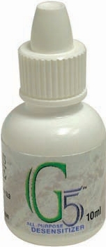
2: After total etching with phosphoric acid, use the antimicrobial agent Gluteraldehyde (G5-Clinical Research Dental (Fig. 4), GLUMA-Heraeus Kulzer, Glue/Sense-Centrix) to rewet the etched surface, because of gluteradehyde’s ability to provide the residual effect of preventing growth of bacteria at the interface between composite and tooth interfaces if microleakage were to occur, without negatively affecting the bond strength.
3: Take advantage of the added clinical benefit that Gluteraldehyde has the ability to decrease post-operative sensitivity. oh
Dr. Leendert (Len) Boksman is Adjunct Clinical Professor at The Schulich School of Medicine and Dentistry at the University of Western Ontario, and he is the Director of Clinical Affairs at Clinical Research Dental, London Ontario. He can be reached atlboksman@clinicalresearchdental.com
Oral Health welcomes this original article.
References
1. Kidd EA , Joyston-Bechal S, Beighton D. Microbiological validation of assessment of caries activity during cavity preparation. Caries Res. 1993; 27(5):402-8.
2. Mertz-Fairhurst EJ, Schuster GS, Williams JE, Fairhurst CW. Clinical progress of sealed and unsealed caries. 1. Depth changes and bacterial counts. J Prosthet Dent 1979;42: 521-526.
3. Jensen OE, Handelman SL. Effect of an autopolymerizing sealant on viability of microflora in occlusal dental caries. Scand J Dent Res 1980;88: 382-388.
4. Boksman L, Carson B. Two year retention and caries rates of UltraSeal XT and FluoroShield light cured pit and fissure sealant. Gen Dent 1998 Mar-Apr; 46(2):184-7.
5. ten Cate JM, van Duinen RN. Hyper-mineralization of dentinal lesions adjacent to glass ionomer cement restorations. JDR 1995Vol 74, 1266-1271.
6. Palenik CJ, Behnen MJ, Setcos JC, Miller Ch. Inhibition of microbial adherence and growth by various glass ionomers in vitro. Dent Mater. 1992 Jan;8(1):16-20.
7. McComb D, Ericson D. Antimicrobial action of new, proprietary lining cements. JDR 1987 Vol 66, 1025-1028.
8. Boksman L, Gratton DR, McCutcheon E, Plotzke OB. Clinical evaluation of a glass ionomer cement as a fissure sealant. Quintessence Int. 1987 Oct; 18(10): 707-9.
9. Kan KC, Messer LB, Messer HH. Variability in cytotoxicity and fluoride release of resin-modified glassionomer cements. JDR 1997 Vo 76, 1502-1507.
10. Brannstom M. Infection beneath composite resin restorations: can it be avoided? Op Dent 1987;12:158-163.
11. Gultz J et al. Antimicrobial activity of cavity disinfectants. Gen Dent Mar-Apr 1999: 187-190.
12. Ergucu Z, Hiller Ka, Schmalz G. Influence of dentin on the effectiveness of antibacterial agents. J Endod. 2005 Feb;31(2):124-9.
13. Ozel E, Kocagoz S, Yurdaguven H, Can Say E. Comparison of antibacterial activity of disinfectant solutions with phosphoric acid. Seq #18 -Microbiology/Immunology and Infection Control, Pharmacology/ Therapeutics and Toxicology, Dental Anesthesiology Research http://iadr.confex.com/iadr/eur04/techprogram/abstract_51221.htm
14. Haak R, Wicht MJ et al. Influence of acid etching or a triclosan composite on the caries micro-flora Abstract #3809 IADR San Diego 2002.
15. Wicht MJ et al. A Triclosan-containing compomer reduces lactobacillus predominant in carious lesions. Dent. Mat 2005; 21:831-836.
16. Settembrini L, Boylan R, Strassler H, Scherer W. A comparison of antimicrobial activity of etchants used for a total etch technique. Op Dent 1997, 22, 84-88.
17. Owens BM, Babu JP. Comparison of antimicrobial effects of cavity pre-treatment Agents. AADR San Antonio 2003 #1636.
18. Ritter AV, Heymann HO, Swift EJ Jr, Perdigao J, Rosa BT. Effects of different re-wetting techniques on dentin shear bond strengths. J Esthet Dent 2000;12(2):85-96.
19. Cobb DS, Reinhardt JW, Vargas MA. Effect of HEMA containing dentin desensitizers on shear bond strength of a resin cement. Am J Dent. 1997 Apr;10(2):62-5
20. Chaconas J, Burgess JO. Shear bond strength of two self-etching adhesives. http://iadr.confex.com/iadr/2002SanDiego/techprogram/abstract_18308.htm
21. Felton D, Bergenholtz, Cox CF. Inhibition of bacterial growth under composite restorations following GLUMA pretreatment. JDR 1989 Vol 68, 491-495.
22. Dondi dall”Orlogogio G, Malferrari S. Desensitization effects of Gluma and Gluma 2000 on hypersensitive dentin. Am J Dent. 1993 Dec;6(6): 283-6.
23. Felton DA, Bergenholtz G, Kanoy BE. Evaluation of the desensitizing effect of Gluma Dentin bond on teeth prepared for complete coverage restorations. Int J Prosthodont 1991 May-Jun;4(3): 292-8.
24. Shupbach P, Lutz F, Finger WJ. Closing of dentinal tubules by Gluma desensitizer. Eur J Oral Sci. 1997 Oct;105(5 Pt 1):414-21.
———
When using dental sealants the possibility of sealing in active decay has been extensively debated in the dental literature
———
In the light of adhesive clinical technique used with composite resins, do these studies have any clinical relevancy?
———
These bacteria might restart the carious process when microleakage occurs and clearly phosphoric acid by itself fulfills that role
———
Since composites do shrink when they polymerize and often display microleakage, we can not allow residual bacteria to remain under a cavity preparation